Mulrecon Color is a web-based DICOM Viewer which accommodates volume exploration of full-color datasets such as the ones made available in the pioneering Visible Human Project.
It is intended to be used as a learning tool in gross anatomy to assist in exploration and comprehension of the three-dimensional visuospatial relations of anatomical structures.
The platform-independent solution with touch functionality allows the application to be utilized with a variety of devices. For instance, large touch screen devices useful for educational purposes can be used as well as virtual reality devices with an internet browser.
The user interface mimics a DICOM viewer used in clinical practice while at the same time providing an easily accessible interface. The technology offers commonplace DICOM functionality including alternate views of the imaging data using various 3D reconstruction techniques such as MPR, VR and MIP.
Consequently, learners can begin navigating and visualizing anatomical structures in and outside of the gross anatomy lab in the same way they will see it throughout their careers.
Link to stand-alone viewer here.
The installation files with source code for the viewer can be downloaded here and instructions for how to load datasets can be found here.
In addition, a demonstrational video is available here.
Dataset cases can be found below.
List of cases
-
Visible Ear
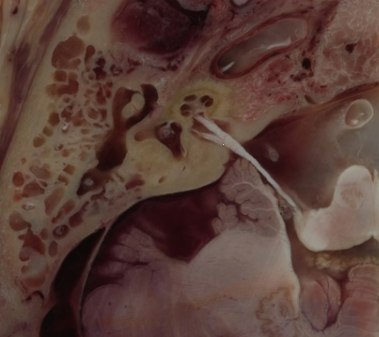
Viewer
Data of dataset: ... Read more
-
Visible Human Male Head
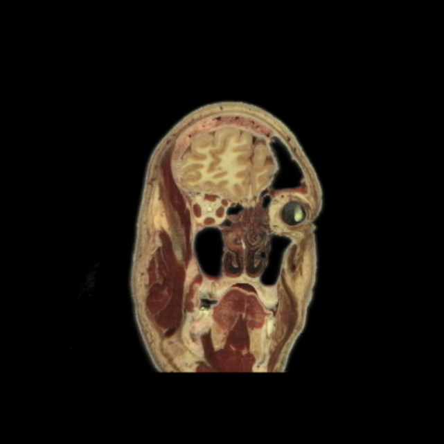
ViewerData of dataset: ... Read more
-
Visible Human Male Head CT
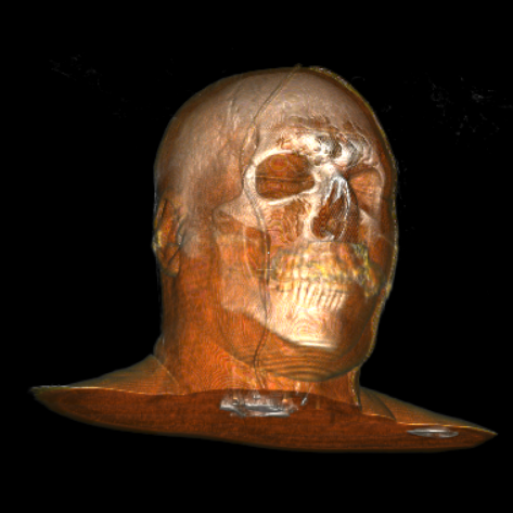
ViewerData of dataset: ... Read more
-
Visible Human Male Head MRI T2
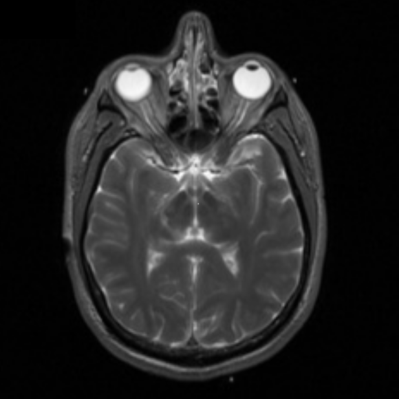
ViewerData of dataset: ... Read more
-
Visible Human 2.0 Head (temporal bone)
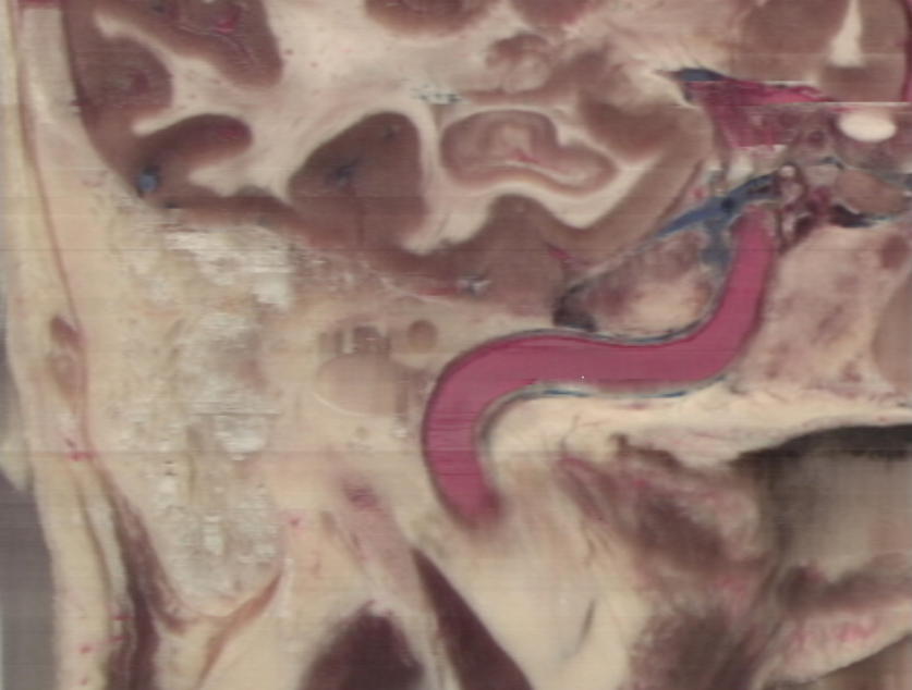
ViewerData of dataset: ... Read more
-
Visible Human 2.0 Head CT
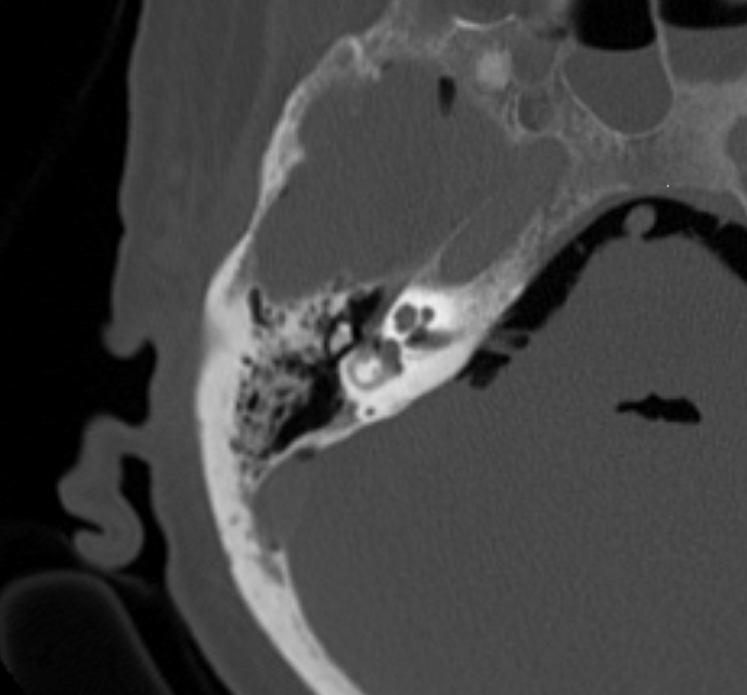
ViewerData of dataset: ... Read more
-
Visible Human Female Hip
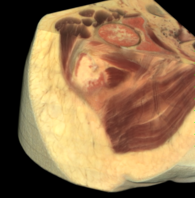
ViewerData of dataset: ... Read more
-
Visible Human Female Hip CT
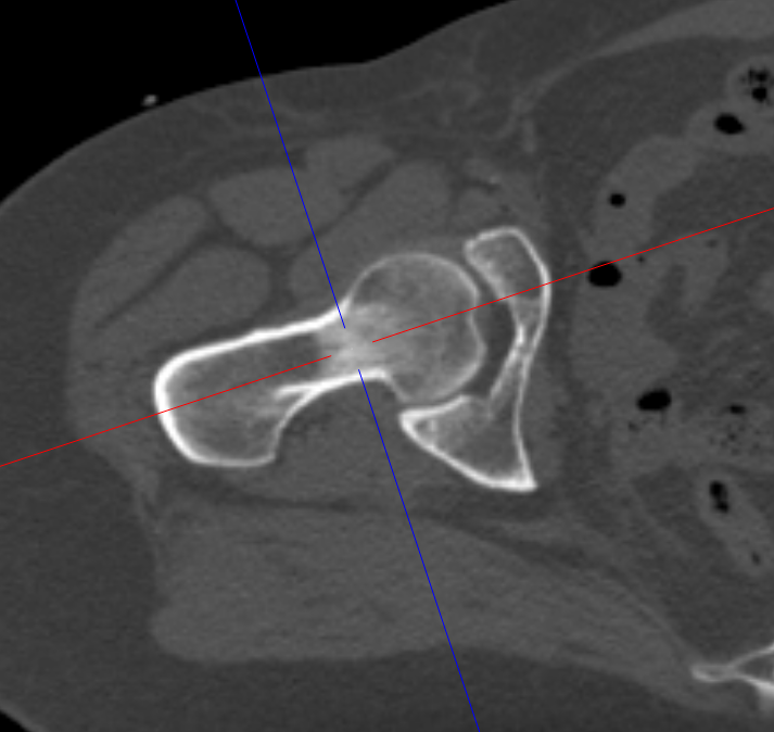
ViewerData of dataset: ... Read more
-
Visible Human Female Hip MRI T2
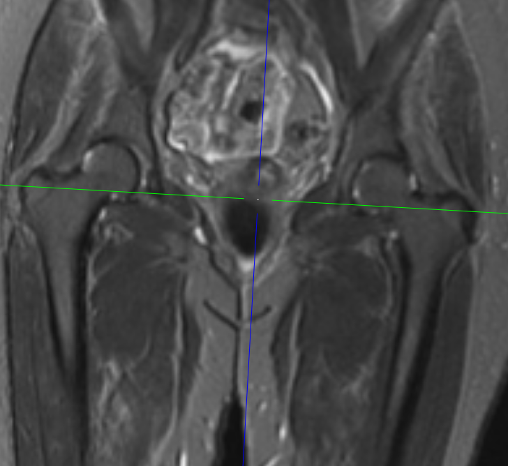
ViewerData of dataset: ... Read more
-
Visible Human Female Ankle
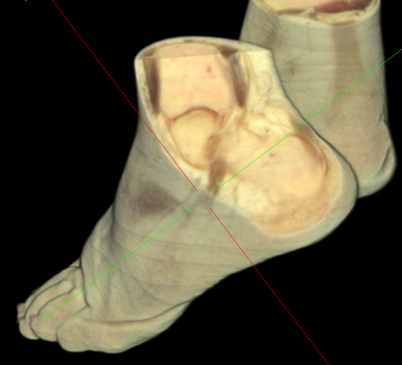
ViewerData of dataset: ... Read more
-
Visible Human Female Ankle CT
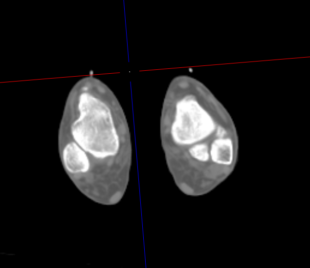
ViewerData of dataset: ... Read more
-
Visible Human Female Eye
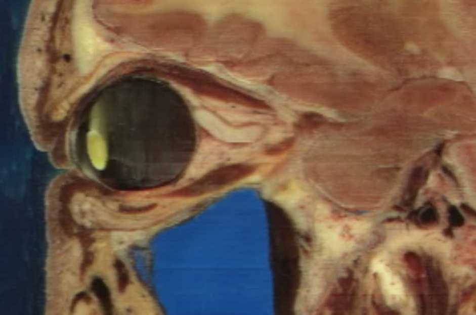
ViewerData of dataset: ... Read more
-
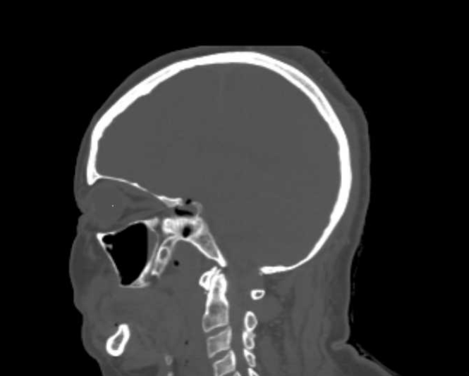
Visible Human Female Eye CT
ViewerData of dataset: ... Read more
-
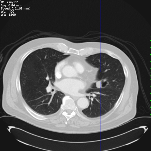
CT thorax of a lung mass in the left lingula
ViewerData of dataset: ... Read more
-
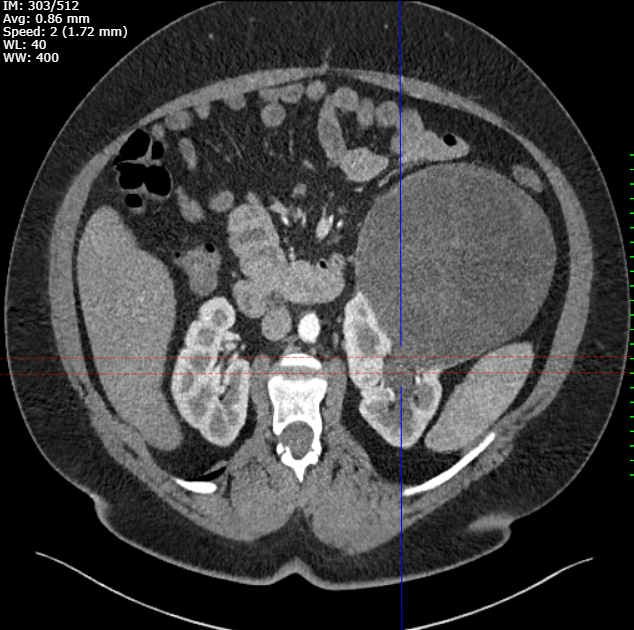
CT abdomen large left-sided renal mass
ViewerData of dataset: ... Read more
-
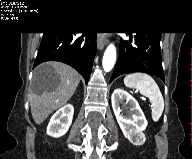
CT abdomen hepatic cysts and left-sided renal mass
ViewerData of dataset: ... Read more
-
MRI T1+contrast of a left frontal lobe mass
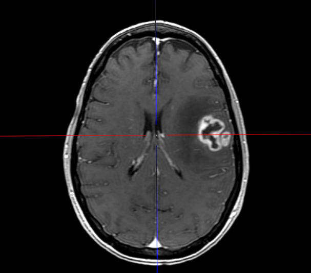
ViewerData of dataset: ... Read more
References
1. The National Library of Medicines Visible Human Project. (2003).
2. Sørensen, M. S. et al. The Visible Ear: A Digital Image Library of the Temporal Bone. ORL vol. 64 378–381 (2002).
3. Clark K, Vendt B, Smith K, Freymann J, Kirby J, Koppel P, Moore S, Phillips S, Maffitt D, Pringle M, Tarbox L, Prior F. The Cancer Imaging Archive (TCIA): Maintaining and Operating a Public Information Repository, Journal of Digital Imaging, Volume 26, Number 6, December, 2013, pp 1045-1057
4. Heller, N., Sathianathen, N., Kalapara, A., Walczak, E., Moore, K., Kaluzniak, H., Rosenberg, J., Blake, P., Rengel, Z., Oestreich, M., Dean, J., Tradewell, M., Shah, A., Tejpaul, R., Edgerton, Z., Peterson, M., Raza, S., Regmi, S., Papanikolopoulos, N., Weight, C. (2019) Data from C4KC-KiTS [Data set]. The Cancer Imaging Archive. DOI: 10.7937/TCIA.2019.IX49E8NX
5. National Cancer Institute Clinical Proteomic Tumor Analysis Consortium (CPTAC). (2018). Radiology Data from the Clinical Proteomic Tumor Analysis Consortium Glioblastoma Multiforme [CPTAC-GBM] collection [Data set]. The Cancer Imaging Archive.